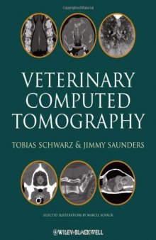
 جزییات کتاب
از زمان تکامل CT scan در سال 1970، بهبود و اصلاحاتی در آن صورت گرفته است و باعث پیشرفت طب بالینی شده است. مشابه رادیوگرافی، CT scan در سال 1890 توسط فیزیکدانان و مهندسین تکامل پیدا کرد اما به سرعت برای احتیاجات طب بالینی تغییراتی در آن صورت گرفت. اولین اسکنرها راه جدیدی برای مشاهده غیر مستقیم مغز و سایر اندام ها که قبلا غیرممکن بود، ارائه دادند. شروع انجام CT scan در دامپزشکی در اواخر دهه 1980 در حیوانات بیمار و در سازمان های پزشکی انسانی انجام گرفت. نصب دستگاه CT scanner در مرکز اسکنر-رادیوتراپی درمحوطه افلورت دامپزشکی بین المللی در پاریس در سال 1980 به عنوان شروع CT scan اختصاصی در دامپزشکی در نظر گرفته می شود. در ابتدا این تکنیک برای تصویربرداری سر سگ و گربه های مبتلا به بیماری های عصبی و بیماری های استخوان بینی مورد استفاده قرار گرفت. با تکامل تکنولوژی حلقه اصطکاک که امکان اسکن مارپیچی را فراهم کرد و هم چنین طراحی دتکتورهای بهتر و کوچکتر امکان اسکن محوطه سینه ای و شش ها و استخوان ها فراهم شد. با پیشرفت تکنولوژی چندین دتکتور در یک ردیف، فصل جدیدی در CT scan دامپزشکی رخ داد. بخش های زیادی از بدن در چندین ثانیه با جزئیات بالا و با کمترین آرتیفکت تصویری، قابل اسکن هستند. CT scan این امکان را برای دامپزشکان فراهم می کند که به طور سریع و با دقت فراوان توانایی تشخیص اختلالات در دام های بزرگ، پستانداران کوچک، پرندگان، خزندگان و حتی حیوانات حیات وحش داشته باشد. هم اکنون CT scan به طور گسترده در حرفه دامپزشکی مورد استفاده قرار می گیرد و قابل مقایسه با سایر روش های تصویربرداری برای بیشتر اختلالات است و دارای پتانسیل گسترده ای به عنوان یک ابزار تشخیصی سریع و موثر برای بسیاری از بیماری ها است.
چندین اطلس و مقاله منتشر شده، CT سگ و بعضی گونه های مشخص را نشان می دهند. به منظور دریافت اطلاعات آناتومیکی جزئی خواننده به این کتاب ارجاع داد ه می شود. بسیاری از اطلاعات علمی منتشر شده در مورد CT scan و بیماری های حیوانات در این کتاب چاپ شده است. این کتاب کمبودی که در مورد مفاهیم تکنولوژیکال، پروتکل های تصویربرداری و ویژگی های CT بیماری های بیشتر گونه های حیوانی در دامپزشکی بود را جبران می کند. بیشتر از 40 دامپزشک متخصص از 13 کشور مختلف، طیف وسیعی از CT در دامپزشکی را در این کتاب کارشناسی کرده اند.
آرزومندیم از خواندن این کتاب راضی باشید و امیدواریم پاسخ برخی سوال های شما در مورد CT دامپزشکی را فراهم کرده باشیم.

Summary by m.mahdavi407
This practical and highly illustrated guide is an essential resource for veterinarians seeking to improve their understanding and use of computed tomography (CT) in practice. It provides a thorough grounding in CT technology, describing the underlying physical principles as well as the different types of scanners. The book also includes principles of CT examination such as guidance on positioning and how to achieve a good image quality. Written by specialists from twelve countries, this book offers a broad range of expertise in veterinary computed tomography, and is the first book to describe the technology, methodology, interpretation principles and CT features of different diseases for most species treated in veterinary practice.Key features• An essential guide for veterinarians using CT in practice• Includes basic principles of CT as well as guidelines on how to carry out an effective examination• Describes CT features of different diseases for most species treated in practice• Written by a range of international leaders in the field• Illustrated with high quality photographs and diagrams throughoutContent: Chapter 1 CT Physics and Instrumentation – Mechanical Design (pages 1–8): Jimmy Saunders and Stefanie OhlerthChapter 2 CT Acquisition Principles (pages 9–27): Tobias Schwarz and Robert O'BrienChapter 3 Principles of CT Image Interpretation (pages 29–34): Jimmy Saunders and Tobias SchwarzChapter 4 Artifacts in CT (pages 35–55): Tobias SchwarzChapter 5 CT Contrast Media and Applications (pages 57–65): Rachel Pollard and Sarah PuchalskiChapter 6 Special Software Applications (pages 67–74): Jennifer Kinns, Robert Malinowski, Fintan McEvoy, Tobias Schwarz and Allison ZwingenbergerChapter 7 Digital Environment (pages 75–76): Robert MalinowskiChapter 8 CT Planning for Radiotherapy (pages 77–80): Lisa J. ForrestChapter 9 Interventional CT (pages 81–87): Tobias Schwarz and Sarah PuchalskiChapter 10 Purchase Considerations (pages 89–92): Victor RendanoChapter 11 Nasal Cavities and Frontal Sinuses (pages 93–109): Jimmy Saunders and Tobias SchwarzChapter 12 Oral Cavity, Mandible, Maxilla and Dental Apparatus (pages 111–124): Lisa J. Forrest and Tobias SchwarzChapter 13 Temporomandibular Joint and Masticatory Apparatus (pages 125–136): Tobias SchwarzChapter 14 Orbita, Salivary Glands and Lacrimal System (pages 137–151): Susanne Boroffka (orbita), Sophie Dennison, Tobias Schwarz and Jimmy SaundersChapter 15 External, Middle and Inner Ear (pages 153–160): Randi DreesChapter 16 Calvarium and Zygomatic Arch (pages 161–170): Federica MorandiChapter 17 Lymph Nodes of Head and Neck (pages 171–174): Olivier TaeymansChapter 18 Pharynx, Larynx and Thyroid Gland (pages 175–184): Olivier Taeymans and Tobias SchwarzChapter 19 Brain (pages 185–195): Silke HechtChapter 20 Pituitary Gland (pages 197–203): Silke Hecht and Tobias SchwarzChapter 21 Cranial Nerves and Associated Skull Foramina (pages 205–208): Laurent CouturierChapter 22 Vertebral Column and Spinal Cord (pages 209–228): Gabriela Seiler, Jennifer Kinns, Sophie Dennison, Jimmy Saunders and Tobias SchwarzChapter 23 Heart and Vessels (pages 229–242): Marc‐André d'Anjou and Tobias SchwarzChapter 24 Trachea (pages 243–248): Tobias Schwarz and Jimmy SaundersChapter 25 Mediastinum (pages 249–260): Audrey Petite and Robert KirbergerChapter 26 Lungs and Bronchi (pages 261–277): Tobias Schwarz and Victoria JohnsonChapter 27 Pleura (pages 279–284): Wilfried MaiChapter 28 Thoracic Boundaries (pages 285–296): Jimmy Saunders, Massimo Vignoli and Ingrid GielenChapter 29 Liver, Gallbladder and Spleen (pages 297–314): Federica Rossi, Federica Morandi and Tobias SchwarzChapter 30 Pancreas (pages 315–324): Ana V. CáceresChapter 31 Gastrointestinal Tract (pages 325–330): Massimo Vignoli and Jimmy SaundersChapter 32 Urinary System (pages 331–338): Tobias SchwarzChapter 33 Genital Tract (pages 339–349): Jimmy Saunders, Federica Rossi and Tobias SchwarzChapter 34 Adrenal Glands (pages 351–355): Federica MorandiChapter 35 Systemic and Portal Abdominal Vasculature (pages 357–370): Tobias SchwarzChapter 36 Abdominal Lymph Nodes and Lymphatic Collecting System (pages 371–379): Federica Rossi, Michail Patsikas and Erik R. WisnerChapter 37 Long Bones (pages 381–386): Ryan M. Schultz and Erik R. WisnerChapter 38 Joints (pages 387–419): Valerie Samii (stifle), Ingrid Gielen (shoulder, elbow, tarsus), Eberhard Ludewig (carpus), William M. Adams (hip), Ingmar Kiefer (carpus), Henri van Bree (shoulder, elbow, tarsus) and Jimmy SaundersChapter 39 Particularities of Equine CT (pages 421–426): Jimmy Saunders, Alastair Nelson and Katrien VanderperrenChapter 40 Equine Sinonasal and Dental (pages 427–442): Jimmy Saunders and Zoe WindleyChapter 41 Equine Calvarium, Brain and Pituitary Gland (pages 443–449): Jennifer Kinns and Russ TuckerChapter 42 Equine Neck and Spine (pages 451–456): Jimmy Saunders and Hendrik‐Jan BergmanChapter 43 Equine Fractures (pages 457–462): Hendrik‐Jan Bergman and Jimmy SaundersChapter 44 Equine Foot (pages 463–471): Sarah PuchalskiChapter 45 Equine Fetlock (pages 473–482): Katrien Vanderperren and Hendrik‐Jan BergmanChapter 46 Equine Upper Limbs (Carpus, Tarsus, Stifle) (pages 483–501): Hendrik‐Jan Bergman and Jimmy SaundersChapter 47 Ruminant and Porcine (pages 503–507): Fintan McEvoy and Stefanie OhlerthChapter 48 Rabbits and Rodents (pages 509–516): Randi DreesChapter 49 Avian (pages 517–532): Michaela GumpenbergerChapter 50 Chelonians (pages 533–544): Michaela Gumpenberger
 دانلود کتاب
 جزییات کتاب


 این کتاب رو مطالعه کردید؟ نظر شما چیست؟
