 جزییات کتاب
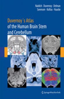
Advanced MRI requires advanced knowledge of anatomy. This volume correlates thin-section brain anatomy with corresponding clinical 3 T MR images in axial, coronal and sagittal planes to demonstrate the anatomic bases for advanced MR imaging. It specifically correlates advanced neuromelanin imaging, susceptibility-weighted imaging, and diffusion tensor tractography with clinical 3 and 4 T MRI to illustrate the precise nuclear and fiber tract anatomy imaged by these techniques. Each region of the brain stem is then analyzed with 9.4 T MRI to show the anatomy of the medulla, pons, midbrain, and portions of the diencephalonin with an in-plane resolution comparable to myelin- and Nissl-stained light microscopy (40-60 microns). The volume is carefully organized as a teaching text, using concise drawings and beautiful anatomic/MRI images to present the information in sequentially finer detail, so the reader easily assimilates the relationships among the structures shown by high-field MRI.


 دانلود کتاب

 جزییات کتاب


 این کتاب رو مطالعه کردید؟ نظر شما چیست؟
